Skin anatomy, illustration - Stock-Grafiken
Illustration of a cross-section through human skin. At bottom are adipocyte (fat) cells (red). Above this is the dermis (flattened yellow cells), and then the basal, spinous and granular layers of the epidermis and keratinised dead cells at the surface. Also shown are fibroblasts (orange), melanocytes (green), langerhans cells (pink) and merkel cells (blue).

Sichern Sie sich dieses Bild in einem von vielen Rahmen auf Photos.com.
EINE LIZENZ KAUFEN
Alle Lizenzen für lizenzfreie Inhalte beinhalten weltweite Nutzungsrechte, umfangreichen Schutz und eine einfache Preisgestaltung mit Mengenrabatten
335,00 €
EUR
Getty ImagesSkin Anatomy Illustration, Stock-Foto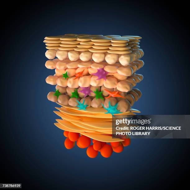 Laden Sie authentische Premium-Grafiken zum Thema Skin anatomy, illustration von Getty Images herunter. Entdecken Sie ähnliche hochauflösende Grafiken in unserem umfangreichen Bildkatalog.Product #:738786939
Laden Sie authentische Premium-Grafiken zum Thema Skin anatomy, illustration von Getty Images herunter. Entdecken Sie ähnliche hochauflösende Grafiken in unserem umfangreichen Bildkatalog.Product #:738786939335€50€
Getty Images
In stockDETAILS
Bildnachweis:
Creative #:
738786939
Lizenztyp:
Kollektion:
Science Photo Library
Max. Dateigröße:
5100 x 5100 px (43,18 x 43,18 cm) - 300 dpi - 5 MB
Hochgeladen am:
Releaseangaben:
Keine Freigaben erforderlich
- Haut,
- Wissenschaft,
- Querschnitt,
- Anatomie,
- Illustration,
- Fettgewebezelle,
- Basalzelle,
- Dreidimensional,
- Langerhanssche Insel,
- Menschliche Haut,
- Bauwerk,
- Biologie,
- Biomedizinische Illustration,
- Dermis,
- Digital generiert,
- Farbbild,
- Fibroblast,
- Gesundheitswesen und Medizin,
- Haut-Pigmentzelle,
- Inneres Organ eines Menschen,
- Kunstwerk,
- Menschlicher Verdauungstrakt,
- Menschliches Körperteil,
- Niemand,
- Quadratisch - Komposition,