menschliches gehirn abschnitt diagramm mit namen - cerebral cortex stock-grafiken, -clipart, -cartoons und -symbole
human brain with highlighted precentral gyrus, illustration - cerebral cortex stock-grafiken, -clipart, -cartoons und -symbole
cross-section of the spinal cord, illustration - cerebral cortex stock-grafiken, -clipart, -cartoons und -symbole
cross-section of the spinal cord, illustration - cerebral cortex stock-grafiken, -clipart, -cartoons und -symbole
digital illustration of prefrontal cortex of human brain highlighted in green - cerebral cortex stock-grafiken, -clipart, -cartoons und -symbole
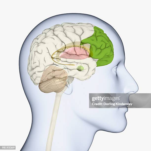
digital illustration of head in profile showing brain of mid adult man with fully developed prefrontal cortex - cerebral cortex stock-grafiken, -clipart, -cartoons und -symbole
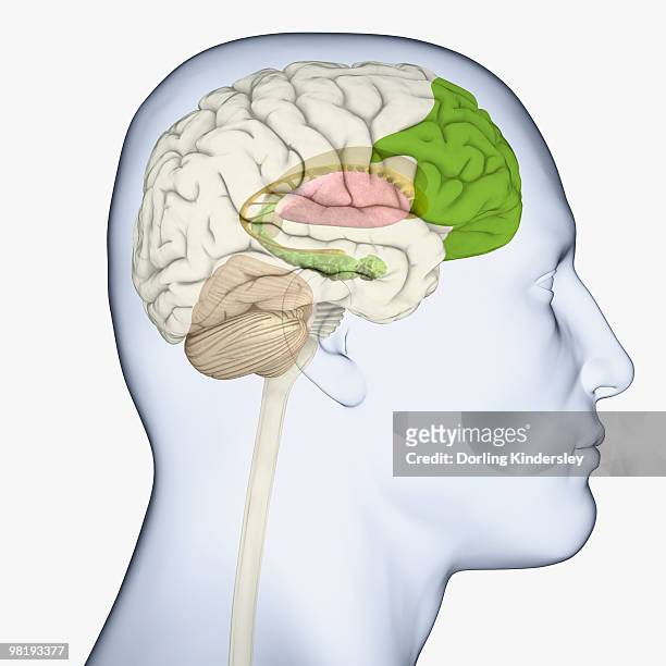
brain with highlighted middle frontal gyrus, illustration - cerebral cortex stock-grafiken, -clipart, -cartoons und -symbole
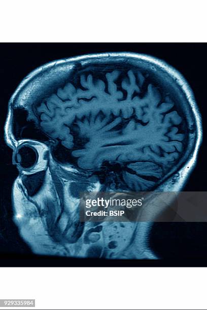
Cerebral atrophy, anterior temporal and parietal frontal Ponto cerebellar, radial cross-section MRI cranial scan.
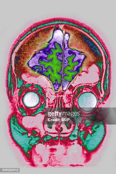
Cerebral atrophy, anterior temporal and parietal frontal Ponto cerebellar, frontal cross-section MRI cranial scan.
Cerebral atrophy, anterior temporal and parietal frontal Ponto cerebellar, frontal cross-section MRI cranial scan.
Cerebral atrophy, anterior temporal and parietal frontal Ponto cerebellar, frontal cross-section MRI cranial scan.
Cerebral atrophy, anterior temporal and parietal frontal Ponto cerebellar, frontal cross-section MRI cranial scan.
Illustration of the location of the black substance, blue). This area contains dopamine neurons that are produced in smaller quantities in the case...
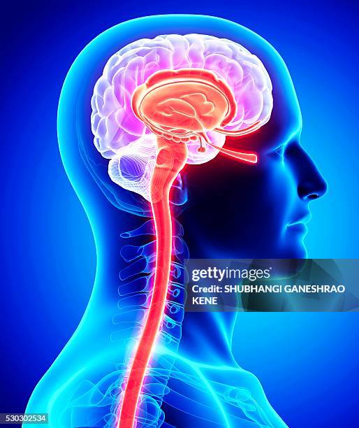
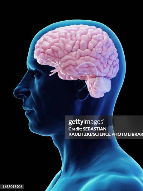
digital illustration of adult human head in profile highlighting parts of brain - cerebral cortex stock-grafiken, -clipart, -cartoons und -symbole
Cerebral atrophy, anterior temporal and parietal frontal Ponto cerebellar, saggital plane MRI cranial scan.
Cerebral atrophy, anterior temporal and parietal frontal Ponto cerebellar, radial cross-section MRI cranial scan.
hippocampal sulcus remnant cysts seen on t2 axial mri (magnetic resonance imaging) - cerebral cortex stock-fotos und bilder
menschliche gehirn anatomie. frontansicht - cerebral cortex stock-grafiken, -clipart, -cartoons und -symbole
digital illustration of anterior part of prefrontal cortex highlighted in green, and direction of dopamine flow in human brain - cerebral cortex stock-grafiken, -clipart, -cartoons und -symbole
vascular malformation in the brain (cavernoma) seen on t2 mri image in the occipital right lobe - cerebral cortex stock-fotos und bilder
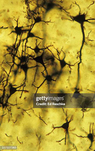
cortex of ranunculus root. parenchyma tissue with starch grains intercellular space. 100x - cerebral cortex stock-fotos und bilder
brain with highlighted middle frontal gyrus, illustration - cerebral cortex stock-grafiken, -clipart, -cartoons und -symbole
digital illustration of reward pathway in human brain - cerebral cortex stock-grafiken, -clipart, -cartoons und -symbole
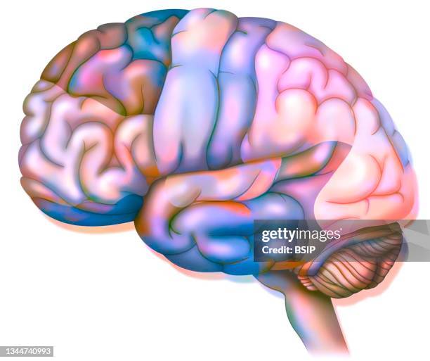
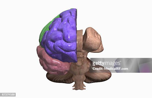
human brain cross-section, illustration - cerebral cortex stock-grafiken, -clipart, -cartoons und -symbole
Illustration of the areas in the brain involved in the process of depression. Lessening in volume and activity of the prefrontal cortex and the...
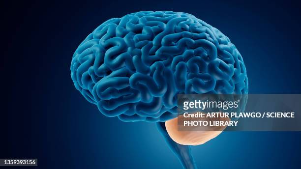
human brain thinking intelligenz symbol und icon - cerebral cortex stock-grafiken, -clipart, -cartoons und -symbole
digital illustration of child's head in profile highlighting parts of brain - cerebral cortex stock-grafiken, -clipart, -cartoons und -symbole
digital illustration of areas of activity during rem sleep in human brain highlighted in red and green - cerebral cortex stock-grafiken, -clipart, -cartoons und -symbole
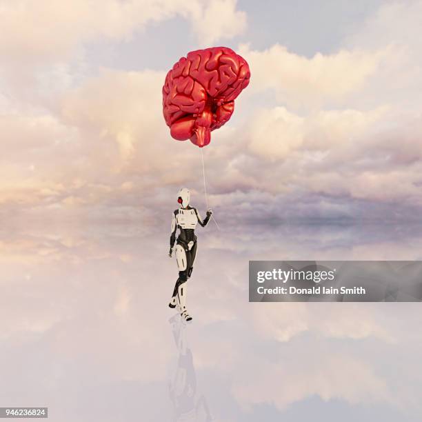
female android holds large floating human brain like balloon on a string - cerebral cortex stock-fotos und bilder
human versus machine: robot supplicates before giant floating human brain - cerebral cortex stock-fotos und bilder
ct scan 84 year old male with alzheimers disease. ct shows brain atrophy with small gyri and large sulci - cerebral cortex stock-fotos und bilder
Whole brain picture information through Magnetic Resoance Imaging MRI. Brain exhibition Inside MIT Museum Building at 265 Massachusetts Avenue...
digital illustration of dorsolateral prefrontal cortex highlighted in green, anterior cingulate cortex and thalamus in human brain - cerebral cortex stock-grafiken, -clipart, -cartoons und -symbole
Cerebral atrophy, anterior temporal and parietal frontal Ponto cerebellar, radial cross-section MRI cranial scan.